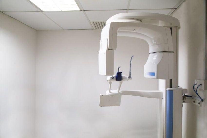
The Panoramic X-ray Department is equipped with a modern digital panoramic X-ray device, for panoramic examination of teeth (in adults and children).
Panoramic imaging of the teeth is a powerful diagnostic tool to evaluate the oral anatomy of a patient (wisdom teeth, temporomandibular joint, jaw bones), while it also reveals the history of previous dental work, such as fillings and root canals.
During the panoramic exam, the dentist detects:
- Problems in the upper and lower jaw of the patient (possible anatomic peculiarities/anomalies).
- Presence of visible tooth decay between the teeth and also in parts not visible to the eye.
- Presence of tooth decay under older fillings.
- The position of dental implants.
- The condition of the remaining bone in the case of periodontal disease.
- Presence of supernumerary teeth.
- Position of impacted wisdom teeth, etc.
The panoramic X-ray is performed with the help of a panoramic X-ray device. It is a short (lasting just a few minutes) and painless exam. During the X-ray, the patient remains standing, while their head is stabilised with a head support. The device then starts rotating around the patient’s head, scanning the area of the upper and lower jaw. After the exam, patients may return to their normal activities.


